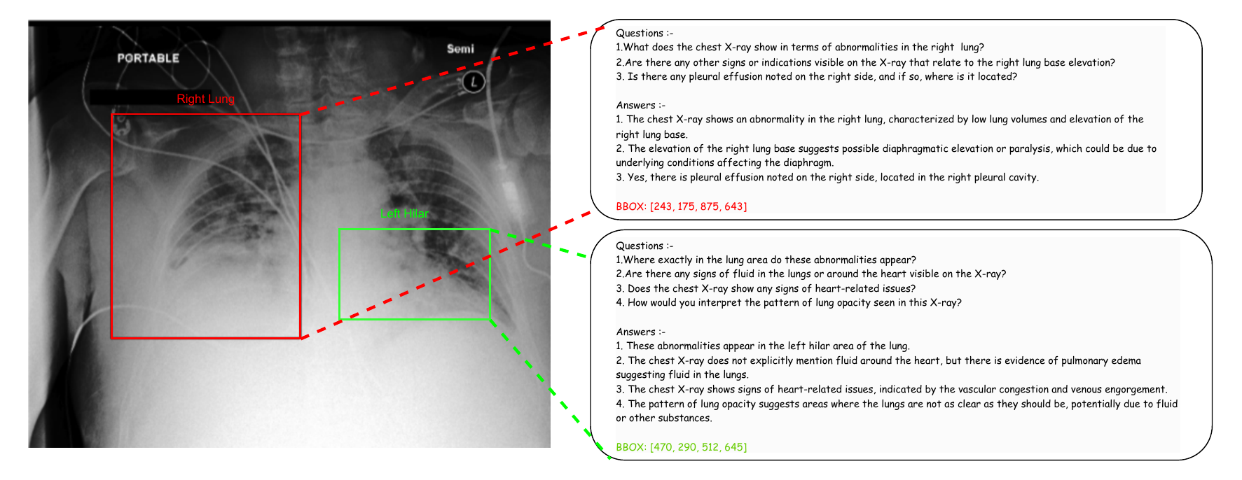
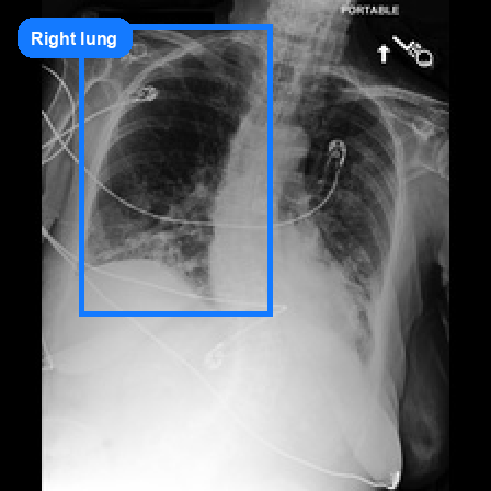
Right Lung
Q: Are there any diseases associated with the lung consolidations and opacity observed?
A: Diseases like pneumonia, pulmonary edema, or lung cancer can be associated with lung consolidations and opacity.
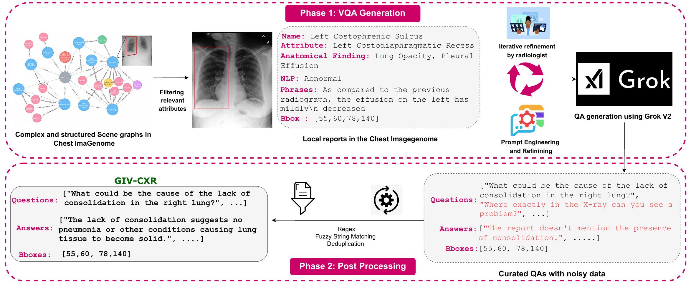
Visual question answering in medical imaging requires models to ground predictions in anatomical locations for clinical verification, yet existing benchmarks lack systematic spatial reasoning evaluation. GIV-CXR introduces a grounded chest X-ray VQA benchmark enabling quantified localization assessment through explicit question-answer to bounding-box linkage and standardized IoU-based evaluation.
The benchmark systematically distributes question-answer pairs across 36 anatomical regions and five reasoning dimensions, combining structured generation, automated hallucination filtering, radiologist validation, and bias analysis to support the development of spatially aware medical vision-language models. We also evaluate pretrained and fine-tuned models including CheXagent, MedGemma-4B, GPT-4o-mini, LLaMA-3.2-11B, and Qwen-2.5-VL variants, showing that explicit spatial supervision substantially improves localization while maintaining strong answer quality.
GIV-CXR is built on MIMIC-CXR and covers abnormality detection, localization, causal reasoning, texture characterization, and differential diagnosis across 36 anatomical structures.
In addition to the dataset itself, the paper benchmarks models such as CheXagent, MedGemma-4B, GPT-4o-mini, LLaMA-3.2-11B, and Qwen-2.5-VL variants to measure answer quality, spatial grounding, clinical validity, and transfer.
We evaluate pretrained multimodal models, fine-tuned baselines, spatial grounding quality, clinical validity, and cross-dataset transfer.
We evaluate CheXagent, MedGemma-4B, GPT-4o-mini, LLaMA-3.2-11B, and Qwen-2.5-VL-7B, along with fine-tuned LLaMA-3.2-11B and two Qwen-based spatial grounding variants: Qwen-BBox-Output and Qwen-BBox-Input.
Answer quality is measured with G-Eval, ROUGE-L, and BERTScore, while spatial grounding is measured with mean IoU and clinical validity is assessed with CheXbert kappa and RadGraph F1.
| Model | G-Eval | ROUGE-L | BERTScore | mIoU |
|---|---|---|---|---|
| CheXagent | 3.22 | 27.05 | 85.12 | - |
| MedGemma-4B | 3.47 | 36.84 | 87.21 | - |
| GPT-4o-mini | 3.13 | - | - | 39.04 |
| LLaMA-3.2-11B (50K) | 3.86 | 70.71 | 92.48 | - |
| Qwen-BBox-Output (150K) | 3.98 | 51.22 | 88.63 | 68.12 |
| Qwen-BBox-Input (150K) | 3.67 | 62.36 | 94.26 | - |
All training, evaluation, and analysis code is available on GitHub. The repo includes fine-tuning notebooks for every model in the paper, evaluation scripts, and bias analysis utilities.
SreevaatsavB / GIV-CXR-CodebaseQuick Start
git clone https://github.com/SreevaatsavB/GIV-CXR-Codebase
cd GIV-CXR-Codebase
python3.11 -m venv .venv && source .venv/bin/activate
pip install -U pip && pip install -e ".[notebooks]"
python scripts/train.py --config configs/qwen_bbox_output.yamlReal samples from the processed release, shown with anatomical regions, questions, answers, and bounding boxes.
Right Lung
Q: Are there any diseases associated with the lung consolidations and opacity observed?
A: Diseases like pneumonia, pulmonary edema, or lung cancer can be associated with lung consolidations and opacity.
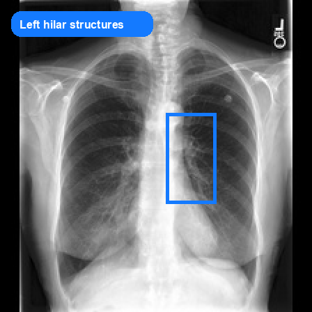
Left Hilar Structures
Q: Which part of the X-ray shows a suspicious opacity in the left hilar region?
A: The left hilar region shows a suspicious opacity.
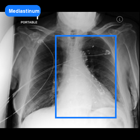
Mediastinum
Q: Can you describe the exact location within the mediastinum where an abnormality is detected?
A: The hiatal hernia is located in the mediastinum, specifically at the level where the stomach herniates through the diaphragm into the chest cavity.
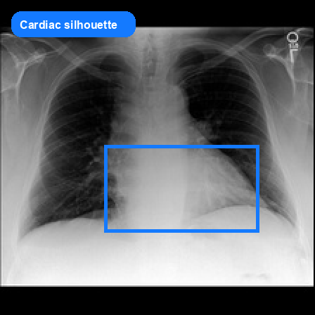
Cardiac Silhouette
Q: Is there anything unusual visible in the heart region from the X-ray?
A: No, the heart region appears normal on the X-ray.
The dataset will be released through PhysioNet under a credentialed data use agreement, consistent with MIMIC-CXR access requirements.
@inproceedings{bavana2026givcxr,
  title     = {GIV-CXR: Densely Grounded, Visually Interpretable Chest X-ray Question Answering Dataset},
  author    = {Bavana, Sreevaatsav and Potta, Adit Rushil and Patnaik, Sai Amrit and Goyal, Nidhi},
  booktitle = {CVPR 2026 Workshop on Medical Reasoning},
  year      = {2026}
}